Busan IFC Mall (2F–4F) · 6,600㎡ One-Day Comprehensive Health Screening & Specialist Care · International Patient Concierge

Medical Guide
Upper GI Series vs. Gastroscopy: Which Test Do You Need
Upper GI series vs gastroscopy: what barium X-ray imaging can show, why endoscopy sees and samples tissue directly, and how to judge which test suits you.
Schedule a ConsultationUpper GI Series vs. Gastroscopy: Which Test Do You Need
Medically reviewed by Dr. Park Kwang-hyuk, Gastroenterology Specialist
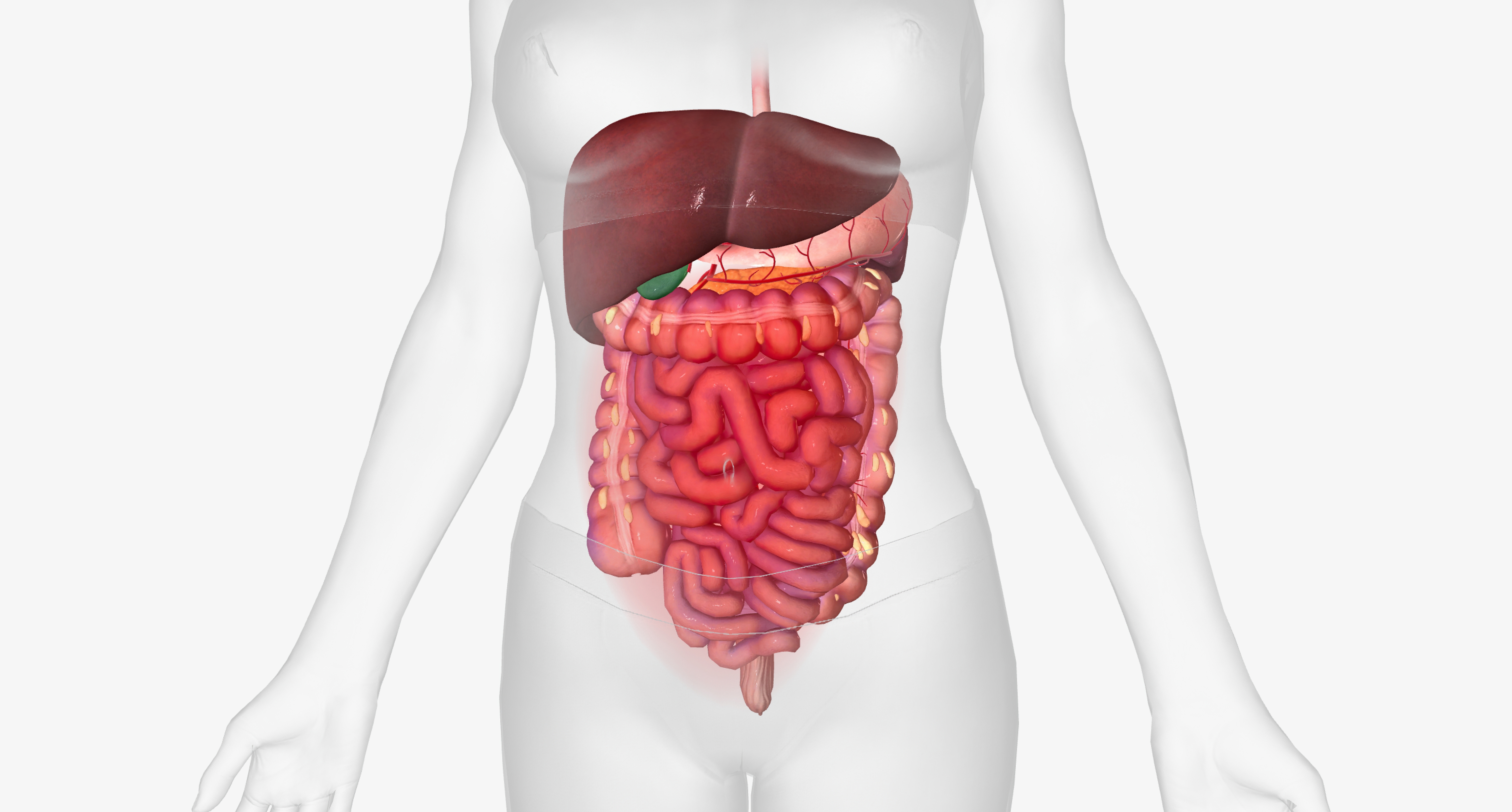
Two examinations can look at the same stretch of your digestive tract, and choosing between them is not a coin flip. An upper GI series vs gastroscopy decision turns on one structural fact: the first produces a picture of a shape, and the second produces a view of a surface. Both are used at Yonsei Mirae IFC Medical Center in Busan, South Korea, and both belong in a screening program. But they answer different questions, carry different risks, and leave you in different places if something looks wrong. This article walks through what each test physically does, what the published comparison research shows, and how to think about the trade-off before you commit to one.
The Bottom Line
- An upper GI series is an X-ray study: you swallow a barium-based contrast agent that coats the esophagus, stomach and duodenum so their outlines appear on fluoroscopic images. It is an indirect examination of structure and function.
- A gastroscopy sends a camera into the same organs, so a physician sees the lining itself, in color, and can take a tissue sample or treat a lesion in the same session.
- The center's own position is that contrast imaging of the upper digestive tract is less accurate than gastroscopy for some findings, which is why an abnormal image often leads to an endoscopy afterwards.
- Population research from Korea's national screening program, published in PLoS One, put the sensitivity of the barium study at roughly half that of endoscopy for gastric cancer detection.
- Neither test finds every abnormality, and neither replaces a conversation about your symptoms, your history and your tolerance for sedation.
What an Upper GI Series Physically Does
The examination has two ingredients: a contrast agent you drink and a moving X-ray. The National Institute of Diabetes and Digestive and Kidney Diseases describes the study as a procedure using X-rays, fluoroscopy and a chalky barium liquid to view the upper digestive tract, in either a standard form using barium alone or a double-contrast form that adds gas-forming crystals to expand the stomach and show its lining in more detail.
Barium is radiopaque, meaning X-rays do not pass through it. When it coats the inner wall of the esophagus, stomach and duodenum, those walls stop being invisible on film and become a traceable outline. A radiologist watches the column of contrast move in real time, which is why the study can comment on function as well as form: how a swallow propagates, whether contrast refluxes back upward, whether something is holding it up. RadiologyInfo.org, the patient resource published jointly by the Radiological Society of North America and the American College of Radiology, lists acid reflux, pain and other symptoms among the reasons the study is ordered, and notes that it is noninvasive.
At Yonsei Mirae IFC Medical Center the sequence is simple to describe. You fast as instructed, drink the prescribed contrast, are positioned for fluoroscopic imaging, and images are captured as the contrast travels down. A physician reviews the images afterwards and advises whether anything requires a further look. The center performs it as a single-visit examination, though the exact time depends on the imaging protocol and on you.
What a Gastroscopy Physically Does
A gastroscopy, also called upper endoscopy or EGD, replaces the shadow with the thing itself. The American Society for Gastrointestinal Endoscopy explains that the examination lets a physician inspect the lining of the esophagus, stomach and duodenum using a thin flexible tube carrying its own light source and camera lens, with the image relayed to a video monitor. The American College of Gastroenterology covers the same examination in its patient material on upper GI endoscopy.
Three things follow from having a camera inside rather than a contrast column outside:
- Color and texture are visible. Inflammation, subtle changes in the mucosal surface, small erosions and early flat lesions have an appearance, not just a shape, and the appearance is what distinguishes them.
- Tissue can be taken. The ASGE notes that biopsy is used both to separate benign from malignant tissue and to test for Helicobacter pylori, the bacterium associated with ulcer disease.
- Treatment can happen immediately. Bleeding can be controlled and certain lesions removed during the same session rather than at a second appointment.
Upper endoscopy in Busan is performed with the GIF-H290 gastroscope running on the Olympus 290-series high-definition platform used throughout the endoscopy unit. Sedation is available for patients who want it, with assessment before the examination, vital-sign monitoring throughout and a monitored recovery period afterwards.
The Difference That Matters Most: Indirect Versus Direct
Describing the barium study as indirect is not a euphemism. You are looking at the negative space a lesion occupies, not at the lesion. A mass large enough to deform the outline of the stomach shows up. A mass that has not yet deformed anything may not. A patch of inflamed mucosa that is perfectly flat has no shadow to cast.
This is the reason the center tells patients plainly that when the images show an abnormality or an area needing closer evaluation, upper endoscopy may be recommended next, and that contrast imaging is less accurate than gastroscopy for some findings. The honest framing of the barium study is that an abnormal result is usually a referral to another test rather than a diagnosis. Even a normal result carries a different weight than a normal endoscopy, because the study's ability to rule things out is limited by what it can see in the first place.
RadiologyInfo.org makes the same point from the radiology side in its discussion of what the examination can and cannot do. An imaging study that maps contours is excellent at contours and constrained everywhere else.
What the Comparison Research Shows
Korea is an unusually good place to study this question, because its national screening program has offered both examinations at scale for decades, and outcomes are linked to a central cancer registry.
A population-based study published in PLoS One analyzed more than 2.6 million people screened with either the upper gastrointestinal series or endoscopy between 2002 and 2005. Sensitivity for gastric cancer was 36.7 percent for the contrast study and 69.0 percent for endoscopy, with specificity essentially identical at about 96 percent for both. Detection rates differed sharply: 0.68 cancers per 1,000 screenings with the barium study against 2.61 per 1,000 with endoscopy. For localized cancer specifically, endoscopy's advantage was statistically significant.
A separate analysis of the same national program, published in the World Journal of Gastroenterology, reported that the probability of detecting gastric cancer was about 2.9 times higher with upper endoscopy than with upper GI X-ray.
Two cautions about reading those numbers. First, endoscopy's 69 percent sensitivity is not 100 percent; a negative endoscopy reduces the probability of disease substantially but does not eliminate it. Second, these figures describe cancer detection in an asymptomatic screening population. If you are being evaluated for a specific symptom, the relevant question is which test answers that symptom's question, and the American College of Radiology maintains Appropriateness Criteria precisely to help clinicians match an imaging study to a clinical scenario rather than choose by habit.
Risk Profiles Are Different, Not Ranked
Neither examination is the safe one and the risky one. They carry unrelated categories of risk.
The barium study
There is no instrument in your body and no sedation, which removes an entire class of complications. What it adds is radiation and a swallowed contrast load. The center lists possible temporary effects including bloating, nausea, constipation and pale or white stools for a few days, with rare complications more likely in people who have swallowing problems or certain gastrointestinal conditions, plus a small amount of radiation exposure. NIDDK identifies constipation from the barium as the most common complication and also lists allergic reaction and intestinal obstruction, advising urgent care for fever, severe abdominal pain, inability to pass gas or no bowel movement within two days.
Suitability narrows in specific situations. The examination may be inappropriate for people with certain swallowing difficulties, suspected obstruction or perforation, significant aspiration risk, or other conditions in which oral contrast could be unsafe. Because X-rays are used, anyone who is or may be pregnant should say so; NIDDK is explicit that X-rays are not recommended during pregnancy.
The endoscopic examination
Here an instrument passes through the throat, and where sedation is used it brings its own considerations. Breathing and blood pressure can be affected by sedative medication in some people, and lingering dizziness afterwards is a recognized reason to extend the recovery period. The center also lists conditions that make a sedated examination less suitable: advanced age in some cases, along with impaired lung function, cardiac or renal disease, cirrhosis, marked obesity, or an acute illness at the time of the appointment. Recognized procedural risks include bleeding, perforation and infection, and the risk profile shifts when a biopsy or a polypectomy is performed. After a sedated examination you rest in a monitored recovery area, and driving is off the table for the day.
What the examination does not involve is ionizing radiation. That distinction matters most for younger patients and for anyone who has already accumulated imaging studies.
Preparation and the Shape of the Day
Both tests require an empty stomach, for the same reason: residue obscures the view. NIDDK advises no food, drink, smoking or gum for eight hours before an upper GI series. At the center, gastroscopy requires eight hours of fasting including water, and the wider pre-checkup guidance asks you to skip alcohol, hard training sessions and punishing work hours in the run-up, with eating stopped once 9 PM has passed.
The practical difference sits after the examination rather than before it. A barium study ends and you walk out; hydration helps clear the contrast. A sedated gastroscopy ends with observation, and the clinic advises avoiding food for at least an hour afterwards, with extra care around eating if a biopsy was taken. If you are visiting Busan on a compressed itinerary, that difference is worth planning for even though it amounts to a few hours.
Who Might Reasonably Choose Which
There is no universal answer, but the reasoning is not mysterious.
- An imaging-based study may suit someone who needs structural and functional assessment of the esophagus, stomach or duodenum, is undergoing a screening program that includes it, or has a reason to avoid instrumentation or sedation.
- Endoscopy is the stronger choice when tissue diagnosis is plausibly needed, when the concern is early or flat mucosal disease, when H. pylori testing is relevant, or when a finding would need treatment anyway.
- If you have swallowing difficulty, an aspiration concern, or a suspected obstruction, the calculus changes, and the physician decides based on your condition rather than on preference.
- If you are or may be pregnant, say so before either test is booked.
At the Busan center, gastroenterology consultations combine your symptoms with examination results and imaging findings before a test is selected, and Dr. Park Kwang-hyuk, a board-certified gastroenterology specialist and graduate of Hanyang University College of Medicine, is among the physicians covering digestive-disease evaluation. The center runs a one-day, one-stop model, so where both an imaging study and an endoscopic examination are indicated, they can often be sequenced within one visit. English-language support is available for international patients, and questions about which examination your program includes can be raised through the contact page before you fly. Background on the center and its specialty areas is on the about page, and patient impressions are collected on the reviews page.
If a finding does need more than a report, the center runs a One-Step referral route: the patient is linked either to the appropriate department in-house or onward to a tertiary general hospital, with structured follow-up management after that handover. That matters for a traveler: the useful question is not only which test to have, but what happens next if it finds something.
Frequently Asked Questions
Is an upper GI series painful?
It is generally not painful. Some people find the barium unpleasant to drink, and temporary bloating, mild abdominal pressure or discomfort from changing position during imaging is common. There is no instrument involved and no sedation.
Can an upper GI series replace a gastroscopy?
Not reliably. The two examinations produce different kinds of information, and the center's stated position is that contrast imaging is less accurate than gastroscopy for some findings. When images show an abnormality, an endoscopy is often the next step rather than an optional extra.
Why can only one of these tests take a biopsy?
Because only one of them puts an instrument in contact with the tissue. The ASGE describes biopsy during upper endoscopy as a routine step that helps separate benign from malignant tissue and can test for Helicobacter pylori. An X-ray study records images from outside the body and has no way to collect a sample.
How much radiation does the barium study involve?
The examination uses X-rays and fluoroscopy, so it carries a small radiation exposure. The medical team keeps the dose as low as is reasonably achievable while still obtaining diagnostic images, and reviews pregnancy status, medications, allergies and relevant conditions beforehand.
What should I do if my stools turn white afterwards?
That is expected. Barium in the digestive tract can lighten stool color for several days. Unless a physician has restricted your fluids, drinking enough helps the contrast pass. Severe abdominal pain, persistent vomiting, significant difficulty swallowing or an inability to pass stool should be reported promptly.
Does a normal result on either test mean nothing is wrong?
No. Both examinations have limits, and the Korean population research shows those limits differ in size. A normal result is meaningful information to be interpreted alongside your symptoms and history, not a guarantee, and persistent or new symptoms deserve another conversation.
Can both be done during one visit to Korea?
Often, yes. The center works on a one-day, one-stop screening model and can sequence examinations within a single appointment block when both are appropriate. Confirm what your selected program includes in advance, since preparation requirements differ between the two.
This article is for general informational purposes and does not constitute individualized medical advice, and it is not a self-diagnosis tool. Screening options, test selection, and the meaning of any result can only be determined through consultation with a qualified physician.
More Articles
Continue reading our clinical guides.
Is a 3.0T MRI Health Screening Safe? Radiation, Contrast
What MRI screening safety really involves: no ionizing radiation, gadolinium contrast risks, implant disclosure, pregnancy advice and claustrophobia help.
Learn More
Low-Dose CT Lung Cancer Screening in Korea: Who Qualifies
Who should consider low-dose CT lung screening in Korea: age and smoking criteria from the USPSTF and ACS, trial evidence, risks, and follow-up planning.
Learn More
Combining a Health Checkup and GI Screening in Korea
How to combine a health checkup and colonoscopy in Korea on one trip: what the shared fast covers, how the day runs, and real limits of a combined visit.
Learn More