Busan IFC Mall (2F–4F) · 6,600㎡ One-Day Comprehensive Health Screening & Specialist Care · International Patient Concierge
Medical Guide
Breast and Thyroid Ultrasound in Korea: A US Guide
What a breast and thyroid ultrasound in Korea involves, how it complements a mammogram, what to bring and disclose, and what a report can really tell you.
Schedule a ConsultationBreast and Thyroid Ultrasound at a Korean Clinic: What US Women Should Know Before Traveling
Medically reviewed by Dr. Seo Jeong-su, M.D., Ph.D., Board Certified Radiologist
If you are booking a breast and thyroid ultrasound in Korea as part of a checkup trip, the appointment itself is likely to be the easiest part of your day. It takes minutes, uses no radiation, and asks almost nothing of you beforehand. The harder part is knowing what the test is for, what it sits alongside, and how to read the piece of paper you carry home. This article walks through the practical side of a combined breast ultrasound and thyroid ultrasound appointment, the reason it complements a mammogram instead of substituting for one, and the things worth telling the imaging team before anyone picks up a probe.
The Bottom Line
- Breast ultrasound and mammography look for different things. Major US bodies treat ultrasound as a supplemental or problem-solving test layered onto mammography, not as a swap for it.
- Dense breast tissue is the single most common reason a supplemental ultrasound comes up, and the National Cancer Institute reports that nearly half of women aged 40 and older who have a mammogram are found to have dense breasts.
- Thyroid nodules are found so often on ultrasound that finding one is closer to a normal result than an alarming one. The American Thyroid Association states that more than 90 percent of thyroid nodules are benign.
- Bring your last mammogram images and report, any prior neck or breast ultrasound, and a written list of medications and surgeries. The value of the study rises sharply when the radiologist can compare against something.
- An ultrasound report describes what was seen and recommends what to do next. It does not, on its own, settle whether tissue is benign or malignant, and no imaging test can exclude cancer.
What the Appointment Actually Involves
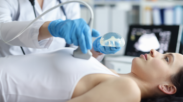
Ultrasound is the imaging method that uses high-frequency sound waves rather than X-rays. As the Radiological Society of North America and the American College of Radiology explain on their joint patient resource RadiologyInfo.org, a small handheld probe called a transducer sends sound into the body, collects the echoes that bounce back, and a computer assembles them into a live picture. There is no ionizing radiation involved, and the images appear in real time, which is why the same technology is used to steer needles during procedures.
The breast portion
You will be asked to undress from the waist up and put on a gown, then lie on your back, usually with the arm on the side being examined raised above your head. That position flattens the breast tissue against the chest wall and gives the probe an even surface to work across. Warm gel goes on the skin so the sound waves can pass through without an air gap, and the examiner sweeps the probe in overlapping passes across each quadrant of the breast and into the armpit.
If you have a lump you can feel, say so and point to it before the scan starts, rather than afterward. The examiner will scan that exact spot deliberately and often measure it in two or three planes. RadiologyInfo.org notes that breast ultrasound is routinely used both to evaluate an abnormality found on a physical exam and to characterize something already seen on a mammogram or breast MRI, and that Doppler settings can be used to assess blood flow within a mass.
The thyroid portion
For the thyroid, you lie on your back with a pillow or wedge under your shoulders so the neck tips gently backward. This feels slightly awkward and is over quickly. The thyroid sits low in the front of the neck, just above the collarbones, in two lobes joined across the middle. The examiner scans both lobes, measures the gland, and records the size, shape, position and internal appearance of any nodule found.
You may be asked to swallow at one point. That is deliberate, because swallowing lifts the gland and lets the examiner see the lower edges of each lobe.
How long it takes
A focused ultrasound of one region is a short study. A combined breast and thyroid examination is still measured in minutes rather than hours, and the largest part of the elapsed time is usually changing, positioning and waiting rather than scanning. At Yonsei Mirae IFC Medical Center in Busan, the Breast & Thyroid Ultrasound service is normally performed within a wider one-day checkup visit, so the realistic planning figure is the length of the whole program you selected rather than the scan alone. A comprehensive checkup at the center is organized as a single visit of roughly 3.5 to 4 hours.
What to Wear and What to Bring
Clothing and same-day preparation
- A two-piece outfit. A top that comes off independently of the bottom half makes both the breast and neck portions simpler.
- No necklace, and ideally no earrings that dangle into the scan field.
- Skip scarves and high collars on the day. The neck needs to be accessible from collarbone to jawline.
- Hair long enough to fall across the throat is best tied back.
- Fasting is not required for breast, thyroid, carotid or certain other ultrasound examinations at the center. If your visit also includes blood work, endoscopy or abdominal imaging, those tests carry their own preparation rules and those rules take precedence.
Deodorant and powder are a mammography concern rather than an ultrasound one, because residue can mimic calcifications on an X-ray image. If your day includes both studies, leave them off.
Prior imaging and reports worth bringing
This is the part US patients most often underestimate. A radiologist reading your scan in Busan has no access to your American records unless you carry them. Comparison is what turns an ambiguous finding into a stable one.
- Your most recent mammogram report, including the stated breast density category.
- The mammogram images themselves on a disc or a portable drive, not just the summary letter.
- Any previous breast ultrasound or breast MRI report.
- Any previous neck or thyroid ultrasound, particularly if a nodule was measured, since size change over time is meaningful information.
- Recent thyroid blood test results, especially TSH.
- A written medication list, including hormone therapy and any blood thinners.
- Dates and descriptions of prior breast surgery, biopsy, implants, or radiation therapy.
What to disclose before the scan
Say these things out loud at the start, even if you wrote them on the intake form:
- Any lump, thickening, skin change, nipple discharge or focal pain, and how long it has been there.
- Where you are in your menstrual cycle, if that applies to you.
- Pregnancy or breastfeeding status. Ultrasound itself is used during pregnancy and lactation, but it changes which other tests belong in the same visit.
- Family history of breast, ovarian or thyroid cancer, and any known genetic result.
- Prior breast implants or reconstruction, which changes scanning technique.
- Prior neck radiation, which is relevant to thyroid assessment.
- Whether you have been told before that you have dense breasts.
Why Ultrasound Complements Mammography Instead of Replacing It
This is the point most worth carrying home, and it is the reason a supplemental scan is offered rather than an alternative one.
The two tests see different things
Mammography is an X-ray study. It is the method that shows microcalcifications, the tiny mineral deposits that can be the earliest visible sign of some breast cancers, and it is the only modality with decades of randomized screening evidence behind it. Ultrasound sees none of that reliably. What ultrasound does well is tell solid from fluid-filled, define the margins and orientation of a mass, and look at tissue that a mammogram renders as an undifferentiated white field.
Because each modality is blind to part of what the other detects, using them together covers more ground than either alone. That is also the logic behind combining mammography and breast ultrasound within the Breast & Thyroid Care services at the Busan center, where breast ultrasound is positioned as the companion study for dense tissue, tissue asymmetry and masses rather than as a standalone screen. The clinic performs its ultrasound studies on high-resolution equipment including an Aplio 300 system.
What dense breast tissue actually means
The National Cancer Institute describes breast density as the proportion of glandular and fibrous connective tissue relative to fatty tissue, as it appears on a mammogram. The American College of Radiology's BI-RADS framework sorts it into four categories, from almost entirely fatty through to extremely dense, and the NCI notes that roughly 40 percent of women fall into each of the two middle-to-high categories, with about 10 percent at each extreme. Only the two upper categories count as "dense."
Two consequences follow, and the NCI separates them deliberately:
- A reading problem. Dense tissue appears white on a mammogram, and so do many tumors and calcifications. White on white is harder to see, so mammography is less sensitive in dense breasts and callbacks are more common.
- A risk problem. Independently of the reading difficulty, women with dense breasts carry a higher baseline risk of breast cancer than women with fatty breasts.
Importantly, the NCI also states that density cannot be felt, not by you and not by a clinician during an exam. The only way to know is from a mammogram report. That is why the density line on your last report is worth carrying with you.
Where the US evidence bodies currently stand
Two authoritative positions are worth understanding, because they are not in conflict even though they read differently.
The US Preventive Services Task Force, in its 2024 breast cancer screening statement, concluded that the evidence is insufficient to weigh the benefits against the harms of supplemental screening with ultrasound or MRI in women found to have dense breasts on an otherwise negative mammogram. The Task Force was explicit that this "I statement" is not a recommendation against supplemental imaging. It is a statement that the trials needed to settle the question have not yet reported the health outcomes that would settle it.
The American College of Radiology approaches the same question through its Appropriateness Criteria, which rate imaging options by clinical scenario rather than issuing a single population recommendation. In the ACR's supplemental screening document based on breast density, ultrasound is rated as a potentially appropriate supplemental option for average-risk women with heterogeneously dense tissue, while MRI carries the stronger rating for women at high risk regardless of density.
The practical translation for you is simple. Supplemental ultrasound is a reasonable, individualized choice discussed with a clinician who knows your risk profile, not a universal instruction. If you already have a US physician managing your breast screening, the sensible move is to decide with them before you travel, not after.
What ultrasound cannot do
It cannot exclude cancer. No imaging test can. Ultrasound can describe a finding as having features that are characteristically benign, or as having features that warrant tissue sampling, and it can document that something has not changed across years. Those are genuinely useful outputs. They are not the same as a negative answer, and any material that suggests otherwise is overselling the technology.
Thyroid Nodules: Why the Finding Itself Needs Context
The thyroid half of the appointment carries a specific risk of unnecessary worry, so it deserves plain numbers.
RadiologyInfo.org states that roughly 5 to 10 percent of adults have a thyroid lump a physician can feel on examination, but that ultrasound is sensitive enough to reveal nodules that cannot be felt, and in some age groups nodules turn up on ultrasound in as many as 70 percent of adults. The overwhelming majority are ordinary variations in thyroid tissue that carry no health consequence. The American Thyroid Association puts the same point in a single figure: more than 90 percent of thyroid nodules are benign.
So the appearance of a nodule on your report is not the signal. What matters is the description attached to it. A radiologist is assessing composition, whether the nodule is solid or fluid-filled, its shape and orientation, the character of its margin, the presence of certain patterns of calcification, and its size. Those features, taken together with your risk factors, determine whether the recommendation is periodic follow-up imaging or sampling with a fine needle. The American Thyroid Association's management guidelines for adults with thyroid nodules set out that reasoning in detail, and note that nodules judged benign or too small to sample are typically monitored with repeat ultrasound over intervals of months rather than acted on immediately.
One further limitation is easy to miss. As RadiologyInfo.org points out, ultrasound shows the structure of the gland but says nothing about whether it is working properly. Function is a blood test question. The National Institute of Diabetes and Digestive and Kidney Diseases describes TSH as the usual first-line blood test for thyroid function, with free T4 and related tests added as needed. This is why thyroid evaluation at the Busan center pairs ultrasound of the gland with TSH, Free T4 and T3 testing: the two answer different questions and neither substitutes for the other.
What Your Report Will and Will Not Tell You
RadiologyInfo.org describes the standard chain: a radiologist interprets the images and issues a signed report to the physician who ordered the study, and the physician goes through the findings with the patient. At Yonsei Mirae IFC Medical Center, international patients can request reports, doctor's notes and medical records in English, and result consultations can also be handled by phone after the visit, which matters when you are reading a report from a different time zone.
Expect the report to contain:
- A description of what was scanned and how.
- Measurements of any discrete finding, in millimeters, with its location.
- A characterization of that finding's features.
- For breast imaging, an assessment category drawn from the American College of Radiology's BI-RADS system, which standardizes both the language and the management recommendation attached to it.
- A recommendation: routine interval, short-interval follow-up, additional imaging, or tissue sampling.
Do not expect it to contain a diagnosis of the tissue itself. That comes only from pathology, which is a separate process after a separate procedure. Do not expect it to reconcile itself with a report you did not provide, and do not expect it to interpret your overall risk. If a finding does require more than imaging, the center connects patients through a One-Step referral pathway to relevant departments or to higher-level general hospitals.
Two practical requests are worth making at the time of the appointment rather than later:
- Ask for the images on physical media as well as the written report, since your physician at home may want their own radiologist to look at them.
- Ask that measurements and any assessment category be stated explicitly in the English report, so a comparison in twelve months has something concrete to compare against.
Practical Notes for a Visit to Busan
Yonsei Mirae IFC Medical Center occupies the second through fourth floors of IFC Mall at 133 Jeonpo-daero in Nam-gu, Busan, next to the BIFC subway station. The facility covers approximately 6,600 square meters and is laid out with separate male and female checkup routes, which means the breast and thyroid portion of a women's checkup is conducted within a female patient flow rather than a shared corridor. For a first-time international patient that detail tends to matter more in practice than it does on paper.
Health screening runs Monday through Friday from 7:30 AM to 4:30 PM and Saturday from 7:30 AM to 12:30 PM, Korean Standard Time, with the clinic closed Sundays and public holidays. The center holds Korea's government registration for treating international patients. English-speaking assistance is available through the international patient service from the first consultation onward, and questions about program contents, scheduling and current costs are best settled in advance through the clinic's contact page. Background on the center's history and services is on its about us page.
Because ultrasound requires no anesthesia, no injection and no recovery, there is no medical reason a routine scan alone should constrain your flight schedule. A finding that triggers further testing is a different conversation, and that conversation belongs to the clinician in front of you.
Frequently Asked Questions
Can breast ultrasound replace my mammogram?
No. The two tests detect different kinds of abnormality, and mammography remains the study with the established screening evidence base behind it. Ultrasound is used to add information, particularly where dense tissue limits what a mammogram can show, and to characterize something already detected. Treat it as an addition to your screening plan rather than a substitution.
Do I need to fast before a breast and thyroid ultrasound?
Not for the ultrasound itself. The center states that fasting is not required for breast, thyroid, carotid and certain other ultrasound examinations. If your visit also includes blood tests, an abdominal scan or endoscopy, those components have their own fasting rules and you should follow the instructions given for the full program you booked.
Does the scan hurt?
It should not. Ultrasound is performed on the surface of the skin with gel and a handheld probe, with no needles and no radiation. You may feel firm pressure, and if the probe passes over an area that is already tender, such as a painful lump or a sensitive spot on the neck, that pressure can be briefly uncomfortable. Tell the examiner and they can adjust.
What does it mean if the report says I have a thyroid nodule?
Usually very little on its own. Nodules are extremely common findings on a sensitive scan, and the American Thyroid Association states that over 90 percent are benign. The meaningful content is the description of the nodule's features and the follow-up interval or sampling recommendation attached to it, which is what you should review with a physician.
Should I bring my American mammogram images?
Yes, and bring the actual images rather than only the summary letter. Comparison with prior studies is one of the strongest tools a radiologist has, because a finding that has been unchanged for years is interpreted very differently from one appearing for the first time. Include the report's breast density category as well.
Will I get results in English?
The center prepares reports, doctor's notes and medical records for international patients in English, and result consultations can be conducted by phone. Ask at the time of your appointment for measurements and any assessment category to be written out explicitly, since those specific details are what a physician at home will want when comparing against future imaging.
Can a normal ultrasound tell me I do not have cancer?
No, and you should be cautious of any source that implies it can. Ultrasound can show that a finding has reassuring characteristics or that nothing has changed since a previous study, both of which are useful. Excluding cancer is beyond what any imaging test can do, which is why tissue sampling exists as a separate step when features warrant it.
This article is for general informational purposes and does not constitute individualized medical advice, and it is not a self-diagnosis tool. Screening options, test selection, and the meaning of any result can only be determined through consultation with a qualified physician.
More Articles
Continue reading our clinical guides.
Is a 3.0T MRI Health Screening Safe? Radiation, Contrast
What MRI screening safety really involves: no ionizing radiation, gadolinium contrast risks, implant disclosure, pregnancy advice and claustrophobia help.
Learn More
Low-Dose CT Lung Cancer Screening in Korea: Who Qualifies
Who should consider low-dose CT lung screening in Korea: age and smoking criteria from the USPSTF and ACS, trial evidence, risks, and follow-up planning.
Learn More
Combining a Health Checkup and GI Screening in Korea
How to combine a health checkup and colonoscopy in Korea on one trip: what the shared fast covers, how the day runs, and real limits of a combined visit.
Learn More