Busan IFC Mall (2F–4F) · 6,600㎡ One-Day Comprehensive Health Screening & Specialist Care · International Patient Concierge

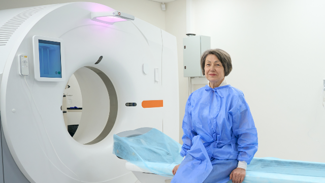
Medical Guide
3.0T MRI Health Screening in Korea vs 1.5T MRI in the US
How a 3.0T MRI differs from a standard 1.5T scanner in signal, detail and trade-offs, and what higher field strength does and does not mean for your scan.
Schedule a Consultation3.0T MRI in a Korean Health Screening: How It Compares With a 1.5T Scanner
Medically reviewed by Dr. Seo Jeong-su, M.D., Ph.D., Board Certified Radiologist
If you are researching MRI health screening Korea options from the United States, one number tends to dominate the marketing copy: the field strength of the scanner. A clinic will say it has a 3.0T MRI, your imaging center at home may run a 1.5T system, and the implication hangs in the air that one is simply better than the other. The honest answer is more interesting than that. Field strength changes the physics of the image in ways that are measurable and real, and it also changes the trade-offs a radiologist has to manage. This guide explains both sides so you can read a scanner specification the way a radiologist reads it.
The Bottom Line
- The "T" in 3.0T stands for tesla, the unit used to measure the strength of the scanner's main magnetic field. A 3.0 tesla system runs a main field twice as strong as a 1.5 tesla system.
- Physics literature describes the intrinsic signal-to-noise ratio of an MRI image as roughly proportional to main field strength, so a higher field generally gives the radiologist more signal to work with.
- That extra signal is a budget, not a verdict. It can be spent on finer spatial detail, on thinner image slices, or on a shorter scan time, and the radiologist decides which matters for the question being asked.
- Higher field strength also changes the picture in less flattering ways: certain artifacts become more prominent, radiofrequency energy deposition rises, and implant labeling is field-specific.
- The right scanner is the one suited to the clinical question. For many routine examinations, a well-run 1.5 tesla study answers the question completely, and no field strength turns a screening scan into a guarantee of health.
What "Tesla" Actually Measures
Magnetic resonance imaging does not photograph your anatomy. It listens to it. As the U.S. Food and Drug Administration explains on its MRI resource pages, an MRI scanner uses a strong magnetic field together with radio waves, and the signal in the resulting image comes mainly from protons in the fat and water molecules in your body. The National Institute of Biomedical Imaging and Bioengineering describes the same process in more detail: the main magnet forces those protons into alignment, a pulse of radiofrequency energy knocks them out of alignment, and the scanner detects the energy released as they settle back. Because different tissues release that energy at different rates, the machine can tell one kind of tissue from another.
The tesla figure describes the strength of that main, always-on magnet. A 1.5 tesla scanner has been the general-purpose clinical workhorse in hospitals worldwide for decades. A 3.0 tesla scanner doubles that main field. Both are, in absolute terms, extraordinarily strong magnets. The National Institute of Biomedical Imaging and Bioengineering makes the point vividly, noting that the field extends beyond the machine itself and exerts enough force on iron-containing objects to fling a wheelchair across the room. That is why the screening questionnaire you fill out before a scan matters more than any specification on a brochure.
Why a Stronger Field Produces More Signal
The practical consequence of doubling the field is a stronger signal relative to background noise. A review of 3 tesla imaging published in the British Journal of Radiology, available through the National Library of Medicine's PubMed Central archive, describes the intrinsic signal-to-noise ratio in magnetic resonance imaging as approximately proportional to the strength of the main magnetic field. More protons align with a stronger field, and more aligned protons mean a louder signal for the scanner to hear.
Noise, in an MRI image, is the graininess that sits between you and the anatomy. Reduce it and the radiologist sees more. But "sees more" splits into several different choices, and this is where the interesting part begins.
The signal budget and what it buys
Extra signal can be spent in at least three ways, and a scanner cannot spend the same signal twice:
- Finer spatial resolution. Smaller image pixels each collect less signal, so a stronger field can support a finer grid without the image dissolving into noise. This is what people mean when they say a 3.0 tesla scan shows smaller structures.
- Thinner slices. MRI builds cross-sectional images. Thinner slices reduce the blurring that happens when several structures are averaged into one image, which matters for small anatomy such as nerve roots, inner ear structures or small joint surfaces.
- Shorter acquisition time. The same image quality can sometimes be obtained faster, which reduces the chance that motion blurs the study and makes the examination easier to tolerate.
A radiologist chooses the mix before the scan starts, based on what the referring question is. That choice is often more consequential for the final report than the field strength itself.
Where the extra detail tends to matter clinically
Higher field strength has been most consistently useful where the target is small, where contrast between adjacent soft tissues is subtle, or where blood flow is being imaged without injected contrast. Neurologic imaging, small-joint musculoskeletal imaging and non-contrast angiographic sequences are the applications most often cited in the literature as benefiting.
It is worth noting how carefully the peer-reviewed evidence is worded, though. A systematic review indexed in PubMed compared 1.5 tesla and 3 tesla magnetic resonance brain imaging across clinical practice and research and concluded that the published evidence demonstrating a clear, decision-changing clinical advantage for the higher field was limited rather than overwhelming. Better pictures and better outcomes are related, but they are not the same claim, and a careful radiology practice does not conflate them.
The Trade-Offs That Come With a Stronger Field
This is the half of the comparison that marketing copy usually omits. Doubling the field does not simply scale everything up; it changes the character of the image.
Artifacts behave differently
An artifact is something that appears in the image but is not in the patient. Several types become more troublesome as field strength rises. Susceptibility artifacts around metal are more pronounced, which can obscure anatomy near dental work, surgical clips or joint hardware. Research published through PubMed Central on metal artifact reduction at both 1.5 and 3.0 tesla documents how specialized sequences are used to manage exactly this problem. Dielectric effects, which produce uneven brightness across a large body region, are also more prominent at higher field and are a known challenge in abdominal and cardiac imaging. A radiologist working at 3 tesla plans around these effects; they are not eliminated by the magnet.
Radiofrequency energy deposition rises
The radio pulses that generate the signal deposit energy in tissue, measured as the specific absorption rate. That deposition rises steeply with field strength. A comparison of fetal brain imaging at 1.5 and 3.0 tesla, published in the peer-reviewed literature indexed by PubMed Central, examined specific absorption rate alongside image quality precisely because the energy question becomes a limiting factor at higher field. In practice this constrains some sequence settings and can make certain protocols slower at 3 tesla than at 1.5 tesla, which partially offsets the speed advantage.
Implant and device compatibility is field-specific
A medical implant is not simply "MRI safe" in the abstract. Labeling is written for specific conditions, and a device tested and labeled for 1.5 tesla is not automatically acceptable at 3 tesla. The American College of Radiology, which publishes the widely used ACR Manual on MR Safety and a suite of MR safety resources for imaging facilities, frames safe practice around screening, documentation and knowing the exact device involved. RadiologyInfo.org, the patient information resource produced by the Radiological Society of North America together with the American College of Radiology, makes the same point in plain language: many modern pacemakers, neurostimulators, cochlear implants and medication pumps are acceptable for MRI, but the technologist and radiologist must know the exact make and model in order to follow the correct procedure.
If you have any implanted device, the useful thing to carry to an appointment abroad is not a preference about field strength. It is the device card or the manufacturer's documentation.
Comfort is not identical
The scanner is louder at higher field. RadiologyInfo.org notes that the system makes loud tapping and knocking noises and that hearing protection is provided as a matter of course. This is normal and expected, and it is one of the practical reasons the preparation conversation matters.
What a Screening MRI Can and Cannot Settle
Field strength is a property of the scanner. It is not a property of the decision to be screened, and the two get confused often enough that it is worth separating them.
Research on magnetic resonance imaging used broadly in people without symptoms has produced a consistent pattern. A review of whole-body MRI for cancer screening in asymptomatic members of the general population, published in a peer-reviewed journal and archived in PubMed Central, reported high rates of findings that are indeterminate or incidental, meaning findings that require further investigation and usually turn out to be benign. The reviewers were cautious about promoting broad, unselected magnetic resonance screening on that basis. A scan with more detail finds more things, and not all of those things are diseases.
That is not an argument against imaging. It is an argument for choosing imaging around a question. A targeted MRI ordered because of a family history, a specific risk profile or a particular symptom is a different proposition from an unfocused scan of everything. RadiologyInfo.org frames the decision the way most radiologists would: the benefits of an examination should outweigh the risks in your particular situation, and there are also risks in not getting an indicated scan, including delayed diagnosis.
The practical version of this, for a traveler planning a checkup, is simple. Ask what question the MRI is meant to answer, and ask what happens if the answer is ambiguous.
How the 3.0T Scan Fits Into a One-Day Checkup in Busan
Yonsei Mirae IFC Medical Center is a health screening center in Busan, South Korea, occupying approximately 6,600 square meters across the second, third and fourth floors of IFC Mall at 133 Jeonpo-daero in Nam-gu, beside BIFC subway station. The center was founded in 2021 and operates as a licensed Korean medical clinic under the Medical Service Act, and it is a Registered Medical Institution for Attracting Foreign Patients certified by Korea's Ministry of Health and Welfare. You can read more about the center's background on its about us page.
Diagnostic imaging is one of six specialty areas at the center, and 3.0T MRI imaging sits alongside low-dose CT and carotid, cardiac and abdominal ultrasound within it. Imaging studies are read by specialists including Dr. Seo Jeong-su, a Board Certified Radiologist who served as a professor at Ewha Womans University College of Medicine, as a clinical professor in the Department of Health Medicine at Samsung Seoul Hospital, and as a visiting fellow at MD Anderson Cancer Center in the United States.
The patient-facing shape of the examination is straightforward:
- Screening begins with a review of your pre-checkup questionnaire to confirm that a magnetic field examination is appropriate for you, followed by selection of the scanning protocol.
- You change into examination clothing free of metal and remove all metallic accessories.
- You lie on the scanner bed and stay still while the images are acquired. The scan itself typically runs about 30 to 45 minutes depending on the anatomy and the sequences requested.
- No anesthesia or sedation is used for a standard diagnostic MRI, and the examination is minimally uncomfortable.
- There is no recovery period, so you can return to normal activity, including travel plans, immediately afterward.
The MRI is one station within a one-day, one-stop screening system; a comprehensive checkup at the center generally runs about 3.5 to 4 hours in total. Screening programs are tiered from Basic through Silver, Gold, Premium and Platinum, and MRI is among the options that can be included depending on which program you select. What a given program contains, and what it costs, is worth confirming directly with the center when you schedule a consultation.
For international patients, a dedicated international medical center provides a foreign-patient coordinator, English and Chinese interpretation, and examination reports, doctor's notes and medical records prepared in English and Chinese. That last point matters more than most scanner specifications: an imaging study you cannot hand to your physician at home in a readable form is an incomplete investment. If a finding needs more than a report, the center operates a One-Step referral system that connects patients to the relevant department or to a higher-level general hospital, with follow-up management.
Questions Worth Asking Before You Book
- Which body region is the MRI covering, and what specific question is it meant to answer?
- Will intravenous contrast be used, and if so, why?
- Do I have any implanted device, and do I have its documentation with me?
- How will the images and the radiologist's report reach my physician at home?
- If something indeterminate turns up, what is the follow-up pathway while I am still in Korea, and after I fly home?
Frequently Asked Questions
Is a 3.0T MRI always better than a 1.5T MRI?
No. A stronger main magnetic field generally provides more signal, which can be converted into finer detail, thinner slices or a faster scan. It also increases certain artifacts and radiofrequency energy deposition, and implant labeling is specific to field strength. The better scanner is the one suited to the clinical question and the individual patient.
Does a 3.0T MRI expose me to radiation?
No. Magnetic resonance imaging uses a magnetic field and radio waves rather than ionizing radiation, which is one of the ways the National Institute of Biomedical Imaging and Bioengineering distinguishes it from x-ray and computed tomography. That is true at every field strength. Separate safety considerations apply to the magnet itself, to implanted devices and to contrast agents.
How long does the scan take, and do I need sedation?
A targeted 3.0T MRI examination at Yonsei Mirae IFC Medical Center generally takes about 30 to 45 minutes, depending on the anatomy being imaged and the sequences requested. Standard diagnostic scans require no anesthesia or sedative. Your main task is to lie still, because motion blurs the images.
Can I have an MRI if I have a pacemaker or a metal implant?
It depends entirely on the device. Many modern implants are acceptable under specific conditions, but the imaging team must know the exact make and model to follow the correct procedure, and labeling written for 1.5 tesla does not automatically apply at 3 tesla. Bring your device card or manufacturer documentation and disclose everything on the pre-scan questionnaire.
Will a 3.0T MRI find everything?
No imaging study does that. More detailed images reveal more findings, and research on magnetic resonance screening in people without symptoms shows that many of those findings are incidental or indeterminate and need further evaluation. A scan supports a clinical judgment; it does not replace one.
How soon will I get my results, and can I fly home afterward?
Initial findings are available the same day through the center's one-stop diagnostic process, with the full radiologist's report following shortly after. Because no sedation is used for a standard MRI, there is no restriction on travel related to the scan itself. Leaving a buffer day in your itinerary is still sensible in case a finding prompts an additional test.
Do I need to fast before an MRI?
It depends on the body region and on whether contrast is planned. The center provides pre-checkup guidelines that cover fasting duration when contrast or clear organ visualization is required, along with instructions to remove metallic accessories and to complete the safety questionnaire in advance. Follow the instructions you receive rather than a general rule.
This article is for general informational purposes and does not constitute individualized medical advice, and it is not a self-diagnosis tool. Screening options, test selection, and the meaning of any result can only be determined through consultation with a qualified physician.
More Articles
Continue reading our clinical guides.
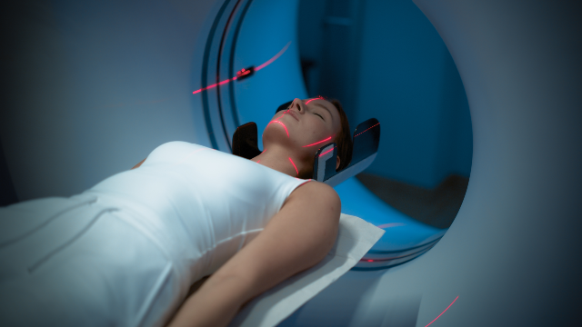
Is a 3.0T MRI Health Screening Safe? Radiation, Contrast
What MRI screening safety really involves: no ionizing radiation, gadolinium contrast risks, implant disclosure, pregnancy advice and claustrophobia help.
Learn More
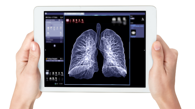
Low-Dose CT Lung Cancer Screening in Korea: Who Qualifies
Who should consider low-dose CT lung screening in Korea: age and smoking criteria from the USPSTF and ACS, trial evidence, risks, and follow-up planning.
Learn More

Combining a Health Checkup and GI Screening in Korea
How to combine a health checkup and colonoscopy in Korea on one trip: what the shared fast covers, how the day runs, and real limits of a combined visit.
Learn More