Busan IFC Mall (2F–4F) · 6,600㎡ One-Day Comprehensive Health Screening & Specialist Care · International Patient Concierge

Diagnostic Imaging · Consultation for Pricing
Carotid, Cardiac & Abdominal Ultrasound
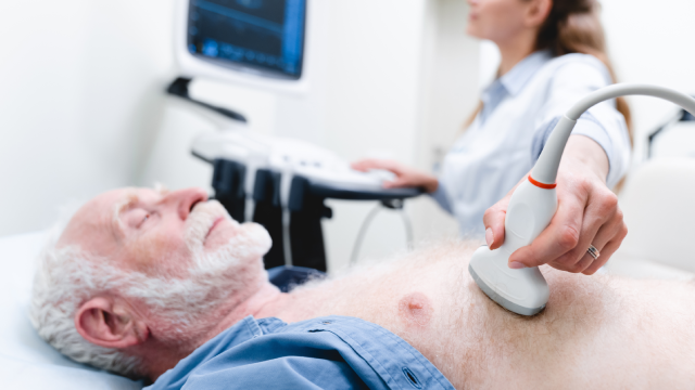
Carotid ultrasound in Busan, cardiac ultrasound and abdominal ultrasound are grouped into one examination set at Yonsei Mirae IFC Medical Center because they share a technology and a purpose: high-frequency sound waves...
Schedule a ConsultationCarotid, Cardiac & Abdominal Ultrasound
Medically reviewed by Dr. Baik Seung Hyuk, M.D., Ph.D., FASCRS, Hospital Director and Medical Director of Yonsei Mirae IFC Medical Center
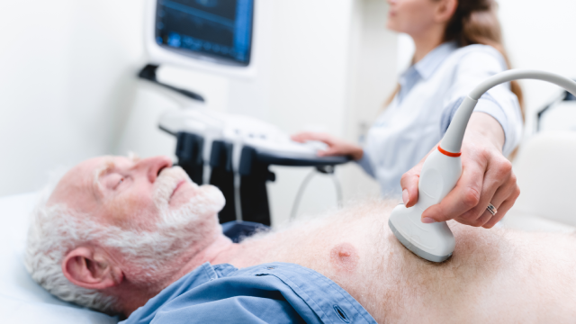
Carotid ultrasound in Busan, cardiac ultrasound and abdominal ultrasound are grouped into one examination set at Yonsei Mirae IFC Medical Center because they share a technology and a purpose: high-frequency sound waves producing real-time images, used to assess the arteries in the neck, the structure and function of the heart, and the organs of the abdomen, without any ionising radiation. This page covers what each of the three assesses, how the visit is organised, and what ultrasound cannot see.
What Is Carotid, Cardiac and Abdominal Ultrasound?
It is three examinations performed with the same modality, each directed at a different region. Ultrasound sends high-frequency sound into tissue and constructs an image from what returns; adding Doppler shows movement, which for blood means flow.
- Carotid ultrasound assesses blood flow in the carotid arteries and the presence of plaque, which relates to stroke risk.
- Cardiac ultrasound assesses the structure and function of the heart, including valve function and pumping efficiency.
- Abdominal ultrasound assesses the appearance of the abdominal organs.
- No ionising radiation is involved in any of the three, and none requires anaesthesia or numbing of any kind.
- The equipment is high-definition Doppler ultrasound with probes selected for vascular flow mapping and for organ structure.
The two halves of what these examinations do — anatomy and flow — are documented together. The National Heart, Lung, and Blood Institute describes carotid ultrasound as using high-frequency sound waves to detect plaque build-up in the carotid arteries and, combined with Doppler, as also showing how blood moves through them, and describes Doppler echocardiography as likewise showing blood flow through the heart's chambers and valves. A picture of a vessel tells you its shape; the Doppler tells you what is happening inside it, which is frequently the more useful of the two.
One further property of these examinations is worth naming early, because it changes what they are for. Because nothing is injected and no radiation is involved, the examinations can be repeated as often as a question requires — which makes them useful as a baseline rather than only as an answer. The clinic frames the results that way: preliminary assessments are generated during the examination itself and establish vascular and organ measurements that a later examination can be compared against. A single carotid scan says what the arteries look like today; two scans several years apart say something about direction, which is usually the more actionable of the two.
Who Performs Carotid, Cardiac and Abdominal Ultrasound at Yonsei Mirae IFC Medical Center
Yonsei Mirae IFC Medical Center documents this service as being overseen by the physician named above rather than documenting that one individual personally performs every appointment, so the member of the clinical team you see on the day is determined by scheduling. Dr. Baik Seung Hyuk, who oversees the general screening team, is a board-certified colorectal surgeon holding M.D. and Ph.D. degrees and an International Fellow of the American Society of Colon and Rectal Surgeons. He was formerly a professor of surgery at Yonsei University College of Medicine's Severance Hospital and director of its Colorectal Cancer Center. Ultrasound is the most operator-dependent of the imaging modalities: unlike a CT or an MRI, which acquire a fixed volume regardless of who presses the button, an ultrasound image exists only where the probe is pointed at the moment of the examination. What the examination finds therefore depends on how it is performed, not only on the equipment performing it.
The Three Examinations Compared
Each region is a different question with a different preparation.
| Examination | What it assesses | Preparation | Typical duration |
|---|---|---|---|
| Carotid ultrasound | Plaque in the carotid arteries and the flow through them; relates to stroke risk | None | About 15–30 minutes |
| Cardiac ultrasound | Heart structure, valve function and pumping efficiency | None | About 15–30 minutes |
| Abdominal ultrasound | The appearance of the abdominal organs | Fasting for approximately 8 hours | About 15–30 minutes |
All three regions can be completed in a single clinic visit, and a combined assessment generally fits within about an hour. Compared with CT or angiography, ultrasound provides real-time soft-tissue and blood-flow evaluation with no radiation and without requiring a contrast injection — which is the reason it is usually the first vascular examination rather than the last.
Who Is a Good Candidate for These Ultrasound Examinations?
- Individuals seeking early detection of vascular plaque.
- People having cardiac valve function or pumping efficiency assessed.
- People having the structure of their abdominal organs evaluated.
- People undergoing routine metabolic health monitoring.
- Individuals seeking a stroke-risk assessment, or screening for liver or kidney disease.
- Patients with extensive open abdominal wounds, active skin infections or unhealed surgical sites across the neck, chest or abdominal scanning regions should postpone the examination until healing has occurred.
What to Expect: Procedure, Recovery, and Results Timeline
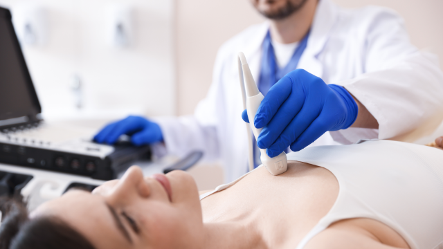
Abdominal ultrasound requires fasting for approximately eight hours beforehand, to reduce gas obstruction and to allow clear imaging of the gallbladder and the other organs. The carotid and cardiac examinations need no preparation. The medical history is reviewed, the appropriate probe settings are selected for each region, the real-time Doppler scans are performed and the findings are interpreted. During the examination the patient lies on an examination bed, warm conductive gel is applied to the skin, and a handheld transducer is moved gently across the neck, the chest and the abdomen. No anaesthesia of any kind is used. Scanning each major region takes roughly fifteen to thirty minutes, so all three can be completed in one visit. There is no recovery period: patients wipe off the gel and resume normal activity, including travel, dining and work. Real-time visual feedback is available during the scan itself, with a same-day preliminary physician evaluation and the formal report issued shortly afterwards.
| Stage | What to expect |
|---|---|
| Fasting | Approximately 8 hours before the abdominal examination; none for carotid or cardiac |
| Position | Lying on an examination bed, with the region being examined exposed |
| The gel | Warm conductive gel applied to the skin before the transducer is moved across it |
| Anaesthesia | None; the examination is entirely non-invasive |
| Duration | Roughly 15–30 minutes per region; a combined assessment within about an hour |
| Sensation | minimally uncomfortable; mild pressure from the transducer, particularly over the abdomen |
| Recovery | None; wipe off the gel and continue the day |
| Results | Real-time feedback during the scan, same-day preliminary evaluation, formal report shortly after |
For international patients this is among the least disruptive examinations available. A stay of only one to two days is typically sufficient, because the examinations are non-invasive and the checkup centre operates a one-day system with fast result processing. Flying home is possible immediately after the examinations and the initial physician consultation, since nothing has been injected, incised or sedated.
Combining These Ultrasound Examinations with Other Imaging
Ultrasound is frequently the anchor of a cardiovascular or metabolic screening programme rather than a standalone examination.
- Carotid, cardiac and abdominal ultrasound are commonly paired with 3.0T MRI where soft-tissue detail elsewhere is also in question.
- They are combined with low-dose CT, which covers the chest that ultrasound cannot image well.
- High-definition gastrointestinal endoscopy is performed alongside them in comprehensive screening programmes.
- The three ultrasound regions are themselves usually performed together, since the preparation for the abdominal examination governs the whole morning.
- Blood testing is routinely run in the same visit, since the fast the abdominal scan requires is the fast most blood panels require anyway.
Each of the three examinations is directed at a specific question — arterial narrowing in the neck, the working of the heart, the appearance of the abdominal organs — and the clinic offers them together as part of detailed health-check programmes rather than as separate bookings. That grouping is what makes a cardiovascular and metabolic overview possible in a morning, and it is also why the results are read against each other rather than one at a time.
Scheduling follows the preparation rather than the other way round. The abdominal examination's eight-hour fast is the constraint that sets the time of the appointment, and because all three regions are normally performed in one sitting, it effectively governs the whole visit — which is why these examinations are usually scheduled first thing in the morning. Where they sit inside a wider checkup, that fast may already be required for the blood tests, in which case nothing extra is being asked. The clinic's facility is organised around this kind of sequencing, with separated movement paths for privacy and automated RFID tracking to keep a multi-examination visit moving between rooms rather than queuing at each one.
Safety, Precision, and Clinic Standards

Diagnostic ultrasound carries no ionising radiation and no documented long-term systemic risk, which places it among the safest imaging modalities available. The side effects are trivial — mild pressure from the transducer during an abdominal scan, and the temporary coldness of the gel. The clinic documents four elements of how it manages quality for these examinations: high-resolution HD ultrasound equipment, expert medical operators, automated RFID workflow tracking and specialised centre pathways. Yonsei Mirae IFC Medical Center operates as a licensed Korean medical clinic under the Medical Service Act.
Safety is not the same as completeness, and ultrasound's limits are physical. RadiologyInfo.org, published by the Radiological Society of North America and the American College of Radiology, describes abdominal ultrasound as imaging the kidneys, liver, gallbladder, bile ducts, pancreas, spleen and abdominal aorta without ionising radiation, with an added Doppler study able to show blockages or reduced blood flow — though sound waves are disrupted by bowel gas and cannot penetrate bone. That is why the eight-hour fast exists: less gas means fewer obscured organs. It is also why a clear ultrasound of the abdomen is not the same statement as a clear CT, and why a symptom that persists after a normal ultrasound may still warrant a different modality.
The clinic describes a One-Step model in which a finding can be routed the same day to an in-house department, or fast-tracked to a partner tertiary hospital.
The International Medical Center at Yonsei Mirae IFC Medical Center provides English-language support for international patients, and the clinic states that this assistance runs from consultation through recovery.
The Bottom Line
- Three radiation-free examinations sharing one technology: carotid arteries, heart, and abdominal organs.
- Doppler is what turns an image of a vessel into information about the blood moving through it.
- The abdominal examination needs an eight-hour fast; the carotid and cardiac examinations need no preparation at all.
- Each region takes roughly 15 to 30 minutes, with no anaesthesia, no recovery and immediate travel afterwards.
- Ultrasound is disrupted by bowel gas and stopped by bone, so a normal result is bounded by what the technique can reach.
Frequently Asked Questions
**Why do I have to fast for eight hours?**Because bowel gas disrupts sound waves and obscures the organs behind it, and because an empty gallbladder images differently from a full one. The fast applies to the abdominal examination specifically; the carotid and cardiac examinations require no preparation.**Does it hurt?**No. The examination is completely non-invasive and minimally uncomfortable. Warm conductive gel is applied and the transducer glides over the skin with light pressure. Some people notice mild pressure during the abdominal portion, and the gel can feel cold at first.**How long will all three take?**Roughly fifteen to thirty minutes per region, so a combined carotid, cardiac and abdominal assessment is generally completed within about an hour. All three fit comfortably into a single clinic visit.**What does the carotid examination actually tell me?**It shows whether plaque has built up in the carotid arteries and, with Doppler, how blood is moving through them. Both relate to stroke risk, which is why this examination frequently appears in cardiovascular screening programmes.**Is there radiation involved?**None. This is the practical difference between ultrasound and CT or angiography: ultrasound provides real-time soft-tissue and blood-flow information with no radiation and without a contrast injection, which is why it can be repeated as often as a question requires.**Can I fly home the same day?**Yes, in general. Nothing is injected, incised or sedated, so patients can travel immediately after completing the examinations and the initial physician consultation. A stay of one to two days is typically sufficient for the whole checkup.**When are the results ready?**Real-time visual feedback is available during the scan itself, so the person performing it is assessing as they go. A same-day preliminary physician evaluation follows, with the formal report issued shortly afterwards.Is there anything that would make me unsuitable?
Extensive open wounds, active skin infections or unhealed surgical sites across the neck, chest or abdominal scanning regions. In those cases the examination is postponed until healing, because the transducer has to make contact with the skin over the area being examined.
Book a Consultation at Yonsei Mirae IFC Medical Center
If the abdominal examination is included, the eight-hour fast decides what time the appointment should be — worth settling when booking rather than afterwards. Enquiries and appointments go through the clinic's enquiry and booking page, or by telephone on . The clinic's ultrasound systems and screening programmes are described further on the About Us page.
Yonsei Mirae IFC Medical Center is located on the second, third and fourth floors of IFC Mall, 133 Jeonpo-daero, Nam-gu, Busan, South Korea. Health screening runs Monday to Friday from 7:30 AM to 4:30 PM and Saturday from 7:30 AM to 12:30 PM, Korean Standard Time. Consultations run Monday to Friday from 8:00 AM to 6:00 PM and Saturday from 8:00 AM to 12:30 PM. The clinic is closed on Sundays and public holidays.
Disclaimer
This page is provided for informational purposes only and does not constitute medical advice, diagnosis or treatment. Whether any test or procedure described here is appropriate for you depends on your own medical history and can only be decided in consultation with a qualified physician.
Sources & Further Reading
- Heart Tests — National Heart, Lung, and Blood Institute, U.S. National Institutes of Health
- Abdominal Ultrasound — Radiological Society of North America and American College of Radiology
Related Medical Services
Explore additional diagnostic examinations and specialist treatments available at Yonsei Mirae IFC Medical Center.
Life-Cycle Checkup Program
The Life-Cycle Checkup Program is a set of age- and life-stage-specific screening packages — pre-marital couples and senior citizens are the two examples the clinic names — designed so that the examinations a person...
Learn More

Fatigue Recovery Clinic
The Fatigue Recovery Clinic at Yonsei Mirae IFC Medical Center provides a structured medical assessment for persistent tiredness or low energy. Fatigue evaluation in Busan here means examining the possible contributing...
Learn More
Upper GI Series
An upper GI series uses swallowed contrast and X-ray imaging to assess structural and functional abnormalities of the oesophagus, stomach and duodenum. The clinic describes it as an indirect examination, in contrast to...
Learn More