Busan IFC Mall (2F–4F) · 6,600㎡ One-Day Comprehensive Health Screening & Specialist Care · International Patient Concierge

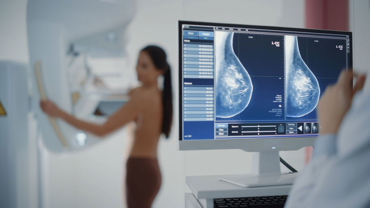
Breast & Thyroid Care · Consultation for Pricing
AI-Assisted 3D Mammography Screening
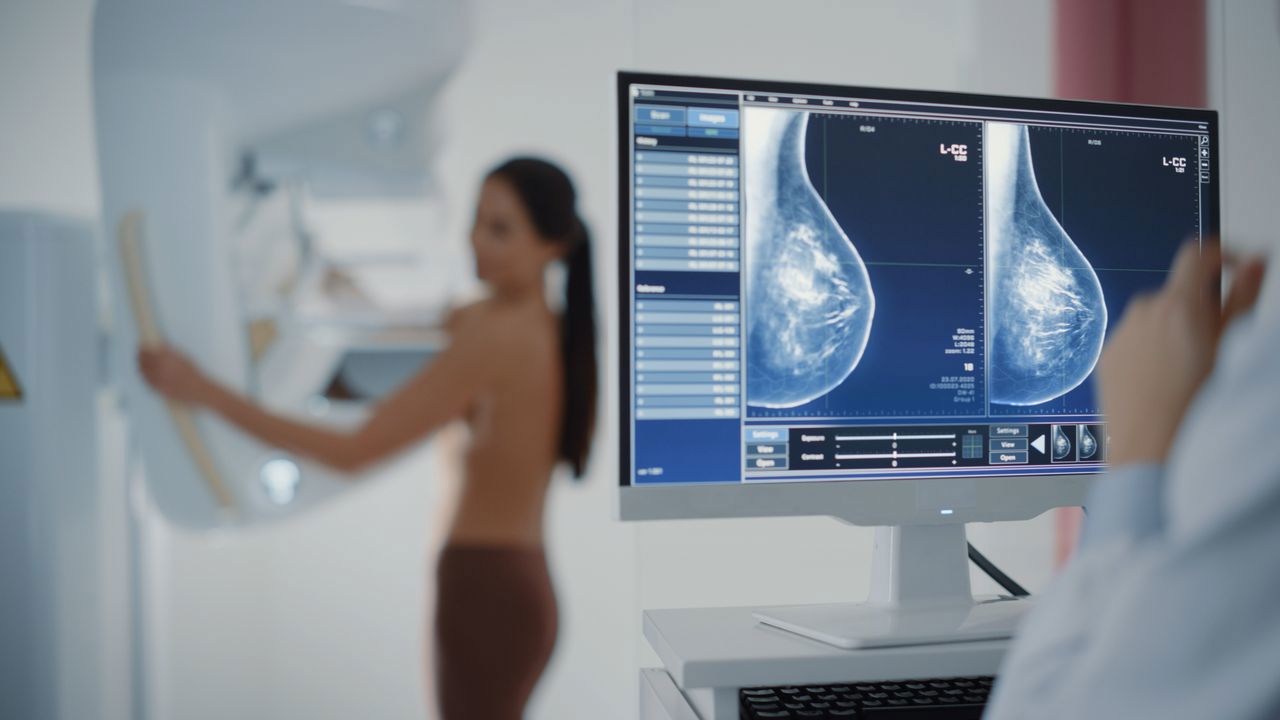
It is a breast X-ray examination performed on a Hologic Selenia Dimensions system with AI-assisted computer-aided detection, used to identify breast abnormalities.
Schedule a ConsultationAI-Assisted 3D Mammography Screening
Medically reviewed by Dr. Kim Seung-il, breast and thyroid specialist and lead of the Breast & Thyroid Center at Yonsei Mirae IFC Medical Center
3D mammography in Busan at Yonsei Mirae IFC Medical Center is an X-ray examination of the breast, supported by AI-assisted computer-aided detection, used to identify abnormalities such as masses, asymmetry and calcifications. The clinic describes mammography as an important examination for detecting suspicious signs of breast cancer. This page covers what the examination involves, the equipment and dose the clinic documents, how to prepare, and the specific circumstances in which mammography alone is not enough.
What Is AI-Assisted 3D Mammography Screening?
It is a breast X-ray examination performed on a Hologic Selenia Dimensions system with AI-assisted computer-aided detection, used to identify breast abnormalities. The AI component supports the assessment of the images; the examination itself is a conventional compression mammogram.
- The examination identifies breast masses, suspicious asymmetry and calcifications — the findings mammography is best placed to show.
- The equipment specification the clinic publishes includes 13.6-million-pixel high-definition imaging, approximately 1.2 mGy radiation exposure, automatic adjustment of the optimal radiation dose, and an ergonomic FAST Paddle.
- Automatic dose adjustment is described as reducing unnecessary exposure, and the high-resolution detector as supporting detailed visualisation.
- Where the breast is dense, or where the mammogram needs additional assessment, breast ultrasound can be added.
- The examination can be performed on its own or within the clinic's comprehensive health-check process, which begins with a questionnaire covering past medical history and current physical condition.
The clinic describes its Hologic Selenia Dimensions mammography system as FDA-approved equipment and states that its AI-based computer-assisted diagnosis is approved by the Ministry of Food and Drug Safety.
What three-dimensional mammography adds is documented in the research literature, with an important qualification attached. The National Cancer Institute reports that digital breast tomosynthesis combined with standard mammography finds more tumours than standard mammography alone, while noting that it is not yet established whether this reduces breast cancer deaths. Both halves of that sentence matter when deciding what to expect from the examination.
Who Performs AI-Assisted 3D Mammography Screening at Yonsei Mirae IFC Medical Center
Yonsei Mirae IFC Medical Center documents this service as being overseen by the physician named above rather than documenting that one individual personally performs every appointment, so the member of the clinical team you see on the day is determined by scheduling. Dr. Kim Seung-il is documented as a breast and thyroid specialist and as the lead of the clinic's Breast & Thyroid Center. The clinic publishes no licence, board certification, education or training details for him, and this page claims none. As with any imaging examination, two distinct people are involved in practice — the radiographer who positions the breast and acquires the images, and the physician who interprets them — and the clinic does not document the positioning staff individually.
Screening Options and Types Available
Mammography is one of several ways of looking at the breast, and the clinic sets out the trade explicitly.
| Examination | What it uses | Where it is strongest | Where it is limited |
|---|---|---|---|
| Mammography | X-rays with compression | Calcifications, suspicious asymmetry, masses | Dense breast tissue can obscure findings |
| Breast ultrasound | Sound waves, no radiation | Dense tissue, characterising masses | Does not show calcifications as mammography does |
| Breast MRI | Magnetic resonance, no radiation | Detailed soft-tissue imaging where clinically indicated | Not a routine screening examination here |
The clinic states plainly that because the lesions visible on each examination differ, complementary testing is most effective — which is why a dense-breast result frequently leads to an ultrasound rather than to a repeat mammogram. The National Cancer Institute notes that breast ultrasound may be offered in addition to mammography for people with dense breasts, but that it is not known whether this supplemental screening leads to better health outcomes.
Who Is a Good Candidate for AI-Assisted 3D Mammography Screening?
- Adults undergoing breast cancer screening, particularly women for whom mammography is appropriate on the basis of age and individual risk.
- Patients being evaluated for a suspicious breast finding rather than screened routinely.
- Women included in the clinic's breast and cervical cancer screening programmes.
- The clinic states that mammography is not performed during breastfeeding; breast ultrasound can be performed during pregnancy or lactation instead.
- Patients with breast implants, previous breast surgery or a previous abnormal mammogram should tell the medical team beforehand.
- Anyone who may be pregnant should raise it before the examination, because the examination uses X-rays.
What to Expect: Procedure, Recovery, and Results Timeline
No major preparation is generally required, with one specific exception: do not apply deodorant, powder, lotion or other products around the breasts or underarms on the day of the examination, because these can create artefacts on the images that resemble calcifications. Tell the medical team about pregnancy concerns, breastfeeding, implants, previous breast surgery or previous abnormal mammograms. No anaesthesia is used. The breast is compressed mechanically to obtain diagnostic images, which can cause temporary pressure or discomfort. Mammography itself is a short examination, though the total time depends on positioning, image acquisition and whether additional views are needed; where it sits inside a comprehensive checkup, the three-and-a-half to four hours that programme takes applies to the whole checkup rather than to the mammogram. No recovery period is normally required and normal activities can be resumed immediately; temporary breast tenderness from compression usually resolves without treatment. Afterwards, patients should receive and review the screening result and follow any recommendation for additional ultrasound, repeat imaging, specialist consultation or other diagnostic work-up.
| Stage | What to expect |
|---|---|
| On the day | No deodorant, powder or lotion on the breasts or underarms — they can mimic calcifications on the images |
| Disclose beforehand | Possible pregnancy, breastfeeding, implants, previous breast surgery, previous abnormal mammogram |
| Anaesthesia | None; the examination is non-invasive |
| During | Mechanical compression of the breast to obtain the images; temporary pressure or discomfort |
| Duration | Short in itself; longer with additional views, and much longer if inside a full checkup |
| Recovery | None; normal activity immediately, with any tenderness resolving on its own |
| Results | Same-day checking within the premium one-day checkup; detailed result consultations by appointment |
| If dense or unclear | Breast ultrasound added for the assessment mammography cannot complete |
For routine mammography alone, an extended stay in Korea is generally not necessary. If a finding calls for additional testing, that changes the schedule rather than the examination.
The consultation around the examination is worth understanding as part of it. Within the clinic's comprehensive health-check process, patients first complete a questionnaire covering past medical history and current physical condition, and the screening examinations are selected on that basis. The mammogram is then performed and interpreted for suspicious breast findings, and the decision about whether ultrasound should be added follows from what the images show rather than being settled in advance. That ordering matters for planning: the possibility of a second examination on the same day is built into how the visit is scheduled.
Combining Mammography with Other Screening
Mammography is rarely the only examination in a visit, and the combinations the clinic documents are specific ones.
- Mammography with breast ultrasound is the strongest documented pairing, particularly where dense breast tissue limits what the mammogram can assess.
- Within the clinic's comprehensive health-check programmes, mammography is bundled alongside cervical screening and HPV testing, body composition measurement, bone-density testing, blood testing and gastrointestinal screening.
- Where breast imaging is part of a one-day checkup, the mammogram is sequenced with the other examinations rather than booked as a separate appointment.
- The clinic's 3.0T MRI can image soft tissue including the breast where that is clinically indicated, though it is not part of routine screening here.
The practical effect for an international patient is that a breast screening examination does not need its own trip: it can be one component of a checkup that answers several questions in the same visit.
Safety, Precision, and Clinic Standards
The principal physical consideration is exposure to a small amount of ionising radiation. The clinic documents its system at approximately 1.2 mGy and describes automatic adjustment of the optimal radiation dose to reduce unnecessary exposure. Mammography does not usually cause medical side effects; the common temporary issue is breast tenderness or discomfort from compression, and there is no incision, injection or anaesthesia involved in the routine examination. The equipment measures the clinic publishes for image quality are the 13.6-million-pixel high-definition detector and the AI-based computer-assisted diagnosis, and for comfort, the ergonomic FAST Paddle.
There are also diagnostic limitations, and the clinic names them rather than leaving them implicit: mammography can produce false-positive and false-negative findings, and dense breast tissue can make some abnormalities harder to detect. That is a property of the examination itself rather than of any particular machine, and it is the reason the dense-breast pathway exists. A normal mammogram is a normal mammogram; it is not a guarantee, and the recommended interval to the next one exists for that reason.
The clinic describes a One-Step model in which a finding can be routed the same day to an in-house department, or fast-tracked to a partner tertiary hospital.
The International Medical Center at Yonsei Mirae IFC Medical Center provides English-language support for international patients, and the clinic states that this assistance runs from consultation through recovery.
The Bottom Line
- The examination is a compression mammogram on a Hologic Selenia Dimensions system with AI-assisted computer-aided detection.
- The documented specification is 13.6-million-pixel imaging, approximately 1.2 mGy, automatic dose adjustment and an ergonomic FAST Paddle.
- Skip deodorant, powder and lotion on the day — they produce artefacts that can look like calcifications.
- Compression causes temporary discomfort; there is no anaesthesia, no recovery period and no restriction afterwards.
- Dense tissue limits what any mammogram can show, which is why breast ultrasound is added rather than the mammogram repeated.
Frequently Asked Questions
**What does the AI component actually do?**It supports the assessment of the images through computer-assisted diagnosis. The examination performed is a conventional compression mammogram; the AI assists with reading it rather than changing what is acquired. The clinic states that its AI-based computer-assisted diagnosis is approved by the Ministry of Food and Drug Safety.**Why can't I wear deodorant?**Because deodorant, powder and lotion around the breasts and underarms can create artefacts on the X-ray images that resemble calcifications. Calcifications are one of the specific findings mammography exists to detect, so an artefact of that kind can mean a repeat examination.**How much radiation will I be exposed to?**The clinic documents approximately 1.2 mGy for its system, with automatic adjustment of the optimal dose to reduce unnecessary exposure. This is a low dose, and it is the reason the examination is performed at an interval rather than routinely repeated.**Will it be painful?**Uncomfortable rather than painful, for most people. Obtaining a diagnostic image requires the breast to be flattened between two plates, and that pressure is what patients notice. It lasts seconds per view. The FAST Paddle on the clinic's system is equipment designed for this part of the examination.**I have dense breasts. Is a mammogram still worth doing?**Yes, but it may not be sufficient on its own. Dense tissue can make some abnormalities harder to detect on a mammogram, which is why the clinic adds breast ultrasound where breast density limits the assessment. The two examinations detect different findings.**Can I have this done while breastfeeding?**No. The clinic states that mammography is not performed during breastfeeding. Breast ultrasound can be performed during pregnancy or lactation, and is the examination used in those circumstances.**When will I get the result?**Within the premium one-day checkup, results can be checked on the same day. Detailed result consultations are available by appointment, and comprehensive checkup result reports are handled through the clinic's own reporting process.What happens if the mammogram shows something?
If the breast is dense or the mammogram requires additional assessment, breast ultrasound can be added. Beyond that, the recommendation may be repeat imaging, a specialist consultation, or further diagnostic work-up, and the clinic's broader screening system provides the follow-up.
Book a Consultation at Yonsei Mirae IFC Medical Center
If you already know your breast tissue is dense, or you have had an abnormal mammogram before, say so when booking — it changes which examinations are scheduled. Enquiries and appointments go through the clinic's enquiry and booking page, or by telephone on . More about the centre, its staffing and its diagnostic infrastructure is on the clinic's About Us page.
Yonsei Mirae IFC Medical Center is located on the second, third and fourth floors of IFC Mall, 133 Jeonpo-daero, Nam-gu, Busan, South Korea. Health screening runs Monday to Friday from 7:30 AM to 4:30 PM and Saturday from 7:30 AM to 12:30 PM, Korean Standard Time. Consultations run Monday to Friday from 8:00 AM to 6:00 PM and Saturday from 8:00 AM to 12:30 PM. The clinic is closed on Sundays and public holidays.
Disclaimer
This page is provided for informational purposes only and does not constitute medical advice, diagnosis or treatment. Whether any test or procedure described here is appropriate for you depends on your own medical history and can only be decided in consultation with a qualified physician.
Sources & Further Reading
- Mammograms — National Cancer Institute, U.S. National Institutes of Health
- Screening for Breast Cancer — National Cancer Institute, U.S. National Institutes of Health
Related Medical Services
Explore additional diagnostic examinations and specialist treatments available at Yonsei Mirae IFC Medical Center.
Life-Cycle Checkup Program
The Life-Cycle Checkup Program is a set of age- and life-stage-specific screening packages — pre-marital couples and senior citizens are the two examples the clinic names — designed so that the examinations a person...
Learn More

Fatigue Recovery Clinic
The Fatigue Recovery Clinic at Yonsei Mirae IFC Medical Center provides a structured medical assessment for persistent tiredness or low energy. Fatigue evaluation in Busan here means examining the possible contributing...
Learn More
Upper GI Series
An upper GI series uses swallowed contrast and X-ray imaging to assess structural and functional abnormalities of the oesophagus, stomach and duodenum. The clinic describes it as an indirect examination, in contrast to...
Learn More