Busan IFC Mall (2F–4F) · 6,600㎡ One-Day Comprehensive Health Screening & Specialist Care · International Patient Concierge

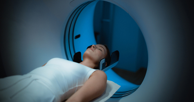
Diagnostic Imaging · Consultation for Pricing
3.0T MRI Imaging
MRI is a non-invasive imaging technique that uses magnetic fields and radio waves to build detailed cross-sectional images.
Schedule a Consultation3.0T MRI Imaging

Medically reviewed by Dr. Seo Jeong-su, board-certified radiologist and diagnostic radiology specialist at Yonsei Mirae IFC Medical Center
A 3.0T MRI scan in Busan at Yonsei Mirae IFC Medical Center uses a strong magnetic field and radio waves — not radiation — to produce high-resolution cross-sectional images of soft tissue, blood vessels and internal organs. The examination itself is undemanding: lie still for thirty to forty-five minutes and it is over. What it asks of a patient beforehand is different, and more important: a complete and honest account of every piece of metal in or on the body. This page covers both halves.
What Is 3.0T MRI Imaging?
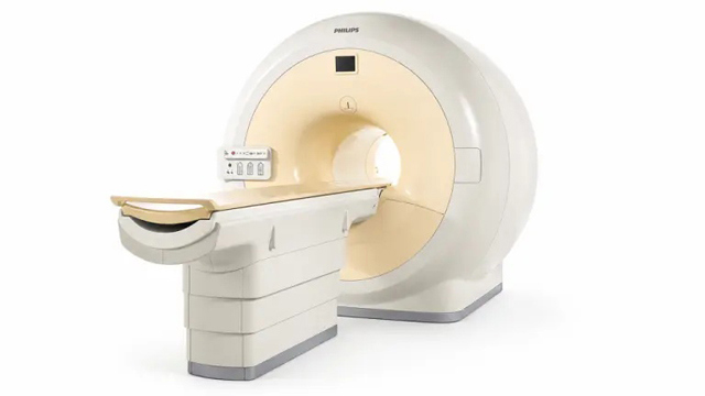
MRI is a non-invasive imaging technique that uses magnetic fields and radio waves to build detailed cross-sectional images. The 3.0T figure refers to the strength of the magnetic field; the clinic describes its system as providing twice the resolution of a traditional 1.5T scanner.
- The scan produces high-resolution images of soft tissue, blood vessels and internal organs, without radiation exposure.
- It is used for detailed neurovascular evaluation, joint and spinal imaging, soft-tissue assessment and comprehensive brain examinations.
- The clinic states that the higher field strength supports clearer visualisation of fine nerve and vascular structures and helps differentiate between tissue types.
- The examination is entirely non-invasive and minimally uncomfortable, and requires no anaesthesia or sedative for a standard diagnostic scan.
- Scanning takes approximately 30 to 45 minutes and is completed within a single clinic visit.
The reason MRI occupies the place it does among imaging options is specific rather than general. The National Institute of Biomedical Imaging and Bioengineering describes MRI as a non-invasive technique that images soft tissue — brain, spinal cord, nerves, muscles, ligaments and tendons — more clearly than x-ray or CT and without ionising radiation, which makes it the modality of choice when repeated imaging is needed. The absence of radiation is what makes MRI repeatable; the soft-tissue clarity is what makes it worth repeating.
Who Performs 3.0T MRI Imaging at Yonsei Mirae IFC Medical Center
Yonsei Mirae IFC Medical Center documents this service as being overseen by the physician named above rather than documenting that one individual personally performs every appointment, so the member of the clinical team you see on the day is determined by scheduling. Dr. Seo Jeong-su is a board-certified radiologist and diagnostic radiology specialist who was formerly a professor at Ewha Womans University College of Medicine, a clinical professor in the department of health medicine at Samsung Seoul Hospital, and a visiting fellow at MD Anderson Cancer Center. An MRI is one examination with two distinct pieces of expertise behind it: choosing and running the scanning protocol, which determines what the images can show, and reading them afterwards. Diagnostic radiology is the specialty that covers both, and it is the specialty documented for this service.
Scan Types and Protocol Options
An MRI is not one examination but a family of them, and the protocol is selected for the question being asked.
| Application | What is imaged | Typical reason |
|---|---|---|
| Brain and neurovascular | Brain tissue and the vessels supplying it | Comprehensive brain assessment, vascular abnormality |
| Spine | Vertebrae, discs and the spinal cord | Spinal symptoms requiring soft-tissue detail |
| Joints | Ligaments, tendons and cartilage | Joint symptoms that X-ray cannot resolve |
| Soft tissue and organs | Organs and the tissue around them | Soft-tissue assessment, including cancer screening |
| MRA (vascular) | Blood vessels specifically | Vascular evaluation alongside a standard sequence |
Compared with a 1.5T MRI or a CT scan, the clinic describes its 3.0T system as providing double the image resolution without ionising radiation. That is an equipment comparison rather than a clinical guarantee: higher field strength resolves smaller structures, which is useful precisely where fine detail is the question, and less decisive where it is not. Screening begins with a pre-checkup questionnaire to confirm safety eligibility, followed by selection of the targeted scanning protocol and same-day physician reporting.
Who Is a Good Candidate for 3.0T MRI Imaging?
- Individuals seeking detailed neurovascular evaluation.
- Patients needing joint or spinal imaging where soft-tissue detail matters.
- Patients undergoing soft-tissue cancer screening.
- Patients having a comprehensive brain examination.
- Patients with non-MR-compatible metallic implants or cardiac pacemakers may not be suitable for a high-field magnetic scan.
- Patients with severe claustrophobia may find the examination difficult, and should raise it before the appointment rather than on the day.
What to Expect: Procedure, Recovery, and Results Timeline
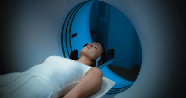
Preparation means removing all metallic accessories, changing into non-metallic examination clothing, and completing a thorough magnetic safety screening questionnaire. On the day the patient completes the safety check, changes into magnet-safe attire, lies on the scanner bed and stays still while the machine acquires the images; the scan finishes in about thirty to forty-five minutes. No anaesthesia or sedation is used for a standard diagnostic scan. The discomforts are acoustic and postural rather than medical: the scanner makes loud tapping noises during acquisition, and lying still for that long can be tiring. Where intravenous contrast is administered, a rare mild allergic reaction is possible. There is no recovery period and no downtime, so normal activities resume immediately afterwards. If contrast was used, drink plenty of fluids afterwards. The images are evaluated through the clinic's streamlined checkup system, with same-day physician reporting.
| Stage | What to expect |
|---|---|
| Before | All metallic accessories removed; magnetic safety screening questionnaire completed |
| Clothing | Non-metallic examination attire provided |
| Position | Lying still on the scanner bed for the duration of the acquisition |
| Duration | Approximately 30 to 45 minutes, within a single visit |
| Sensation | No pain; loud acoustic tapping during acquisition is the main thing patients notice |
| Contrast | Used only where the protocol requires it; rare mild allergic reaction possible |
| Recovery | None; immediate return to normal activity |
| Afterwards | Fluids if contrast was given; same-day physician reporting of the findings |
The facility is organised to make that single visit work: the clinic uses automated RFID tracking and separated movement pathways to manage examination flow, which is what allows a scan of this length to sit inside a one-day programme alongside other examinations.
Combining MRI with Other Examinations
An MRI is frequently one component of a wider imaging plan rather than a standalone appointment, and the clinic documents the combinations it uses.
- MRI is combined with the 128-channel MDCT scanner where a question spans both soft tissue and the chest or bone.
- MRA vascular sequences can be added to a standard MRI protocol where blood vessels are the specific concern.
- Ultrasound is used alongside MRI for organs where real-time imaging and Doppler flow add something the static images do not.
- High-definition endoscopy is combined with imaging in comprehensive organ screening programmes.
Because the MRI is the longest of these examinations, it tends to set the shape of the day when several are combined — which is why it is usually scheduled rather than added on arrival.
Safety, Precision, and Clinic Standards
MRI safety is a screening problem rather than a dose problem, and it is worth understanding why. RadiologyInfo.org, published by the Radiological Society of North America and the American College of Radiology, explains that MRI uses a powerful magnetic field and radio energy rather than ionising radiation, and that the magnetic field can attract ferromagnetic objects or affect implanted devices — which is why every patient must be screened for implants and metal before entering the scanner room. The risk is not cumulative or statistical, as it is with radiation; it is immediate and specific to what a particular person is carrying.
That is the reason the clinic's documented contraindications are what they are: non-MR-compatible metallic implants and cardiac pacemakers. It is also why the safety questionnaire is not a formality to be completed quickly in the waiting room. Surgical clips, stents, cochlear implants, metal fragments from an old injury, and devices a patient may not think of as medical all belong on it. If you are unsure whether something counts, say so rather than deciding for yourself.
The other documented consideration is claustrophobia, which is a genuine reason a scan may be difficult rather than a matter of nerves, and which is better discussed before the appointment than discovered inside the bore. Beyond those, the risks are minor: acoustic noise, the tedium of staying still, and a rare mild allergic reaction where contrast is used. Yonsei Mirae IFC Medical Center operates as a licensed Korean medical clinic under the Medical Service Act.
The clinic describes a One-Step model in which a finding can be routed the same day to an in-house department, or fast-tracked to a partner tertiary hospital.
The International Medical Center at Yonsei Mirae IFC Medical Center provides English-language support for international patients, and the clinic states that this assistance runs from consultation through recovery.
The Bottom Line
- A 3.0T MRI produces high-resolution images of soft tissue, vessels and organs using magnetism and radio waves, with no radiation.
- The scan takes roughly 30 to 45 minutes, needs no anaesthesia and has no recovery period.
- The whole of MRI safety is in the screening: implants, pacemakers and metal must be declared before entering the scanner room.
- Claustrophobia is a documented reason a scan may be unsuitable, and is best raised before the appointment.
- Higher field strength resolves finer detail; it is an equipment property, not a promise about what will be found.
Frequently Asked Questions
**Is there any radiation in an MRI?**None. MRI uses a strong magnetic field and radio waves rather than ionising radiation, which is why it can be repeated when a condition needs following over time, and why it is often preferred where soft tissue is the question.**How long will I be in the scanner?**Approximately 30 to 45 minutes for the scan itself, completed within a single clinic visit. The time varies with the protocol selected, since a scan targeted at one region takes less than one covering several.**What do I need to remove beforehand?**All metallic accessories, and you will change into non-metallic examination clothing. This includes items easily forgotten — hairpins, jewellery, watches, anything with a magnetic strip. The safety screening questionnaire is designed to catch what removing your own accessories will not.**I have a metal implant. Can I still be scanned?**It depends entirely on the implant. Non-MR-compatible metallic implants and cardiac pacemakers are documented contraindications for a high-field magnetic scan. Declare every implant, clip, stent or retained fragment during screening, including ones you assume are irrelevant.**Will it hurt?**No. The examination is entirely non-invasive and minimally uncomfortable and requires no anaesthesia. What patients notice is the loud tapping noise the scanner makes while acquiring images, and the effort of staying still for the duration.**I am claustrophobic. What should I do?**Say so when booking rather than on the day. Severe claustrophobia is documented as a reason a patient may not be suitable for this scan, and knowing in advance allows the appointment to be planned around it instead of abandoned partway through.**Will I need contrast?**Only if the protocol requires it. Where intravenous contrast is used, a rare mild allergic reaction is possible, and the advice afterwards is to drink plenty of fluids. Tell the medical team about any previous reaction to contrast media.When will I know the results?
The scans are evaluated through the clinic's streamlined checkup system with same-day physician reporting, so the findings are normally discussed during the same visit rather than communicated later.
Book a Consultation at Yonsei Mirae IFC Medical Center
If you have an implant, a pacemaker, a history of metal injury or significant claustrophobia, raise it when booking — it decides whether this is the right scan for you. Enquiries and appointments go through the clinic's enquiry and booking page, or by telephone on . The imaging department and its equipment are described further on the clinic's About Us page.
Yonsei Mirae IFC Medical Center is located on the second, third and fourth floors of IFC Mall, 133 Jeonpo-daero, Nam-gu, Busan, South Korea. Health screening runs Monday to Friday from 7:30 AM to 4:30 PM and Saturday from 7:30 AM to 12:30 PM, Korean Standard Time. Consultations run Monday to Friday from 8:00 AM to 6:00 PM and Saturday from 8:00 AM to 12:30 PM. The clinic is closed on Sundays and public holidays.
Disclaimer
This page is provided for informational purposes only and does not constitute medical advice, diagnosis or treatment. Whether any test or procedure described here is appropriate for you depends on your own medical history and can only be decided in consultation with a qualified physician.
Sources & Further Reading
- Magnetic Resonance Imaging (MRI) — National Institute of Biomedical Imaging and Bioengineering, U.S. National Institutes of Health
- MRI Safety — Radiological Society of North America and American College of Radiology
Related Medical Services
Explore additional diagnostic examinations and specialist treatments available at Yonsei Mirae IFC Medical Center.
Life-Cycle Checkup Program
The Life-Cycle Checkup Program is a set of age- and life-stage-specific screening packages — pre-marital couples and senior citizens are the two examples the clinic names — designed so that the examinations a person...
Learn More

Fatigue Recovery Clinic
The Fatigue Recovery Clinic at Yonsei Mirae IFC Medical Center provides a structured medical assessment for persistent tiredness or low energy. Fatigue evaluation in Busan here means examining the possible contributing...
Learn More

Upper GI Series
An upper GI series uses swallowed contrast and X-ray imaging to assess structural and functional abnormalities of the oesophagus, stomach and duodenum. The clinic describes it as an indirect examination, in contrast to...
Learn More